Loculated Pleural Effusion Definition - Pleural Effusion X Ray - slidedocnow - There is normally a tiny amount of fluid between the two layers of pleura.
bymamawhobrey-
0
Loculated Pleural Effusion Definition - Pleural Effusion X Ray - slidedocnow - There is normally a tiny amount of fluid between the two layers of pleura.. Loculated effusions occur most commonly in association with conditions that cause intense pleural inflammation, such as empyema, hemothorax, or tuberculosis. A pleural effusion is accumulation of excessive fluid in the pleural space, the potential space that surrounds each lung. Imaging of pleural plaques, thickening, tumors. Causes of an exudative effusion are it results when the production of pleural fluid exceeds the body's ability to reabsorb it. Pleural effusions can loculate as a result of adhesions.
The lungs and the chest cavity both have a lining that consists of pleura, which is a thin membrane. Causes of an exudative effusion are it results when the production of pleural fluid exceeds the body's ability to reabsorb it. Learn vocabulary, terms and more with flashcards, games and other study tools. A pleural effusion is a collection of fluid next to the lung. Pleural effusion refers to a buildup of fluid in the space between the lungs and the chest cavity.
Loculated pleural effusion | Radiology Case | Radiopaedia.org from images.radiopaedia.org Pleural effusion refers to a buildup of fluid in the space between the lungs and the chest cavity. The effusion, in this case, is restricted to one or more fixed pockets within the pleural space. There is normally a tiny amount of fluid between the two layers of pleura. Compartmentalization of a pleural effusion into smaller spaces by fibrous layers. A pleural effusion is an abnormal buildup of fluid around your lungs, between the layers of tissue that line the lungs and chest cavity. Approximately 1 million people develop this abnormality each year in the most pleural effusions, whether free flowing or loculated, are hypoechoic with a sharp echogenic line that delineates the visceral pleura and lung. Pleural effusion (transudate or exudate) is an accumulation of fluid in the chest or on the lung. Learn about pleural effusion (fluid in the lung) symptoms like shortness of breath and chest pain.
Imaging of pleural plaques, thickening, tumors.
In our study loculated pleural effusion were seen in 8 patients, among which 6 cases were loculated tubercular effusion which were treated with steroids and 2 cases were loculated empyema of which 1had minimal loculations removed by medical thoracoscopy while other had moderate loculations. Encapsulation) is most common when the underlying effusion is due to hemothorax ultrasonography permits easy identification of free or loculated pleural effusions, and it facilitates. The effusion, in this case, is restricted to one or more fixed pockets within the pleural space. Pleural effusion is classically divided into transudate and exudate based on the light criteria. A pleural effusion is a collection of fluid next to the lung. The pleural fluid may loculate between the visceral and parietal pleura (when there is partial fusion of the pleural layers) or within. A pleural effusion is an abnormal buildup of fluid around your lungs, between the layers of tissue that line the lungs and chest cavity. Differentiation of loculated effusions from solid. The body produces pleural fluid in small amounts to lubricate the surfaces of the pleura. The effusion may cause you to become breathless. Left pleural effusion developed 4 days after antibiotic treatment for pneumococcal pneumonia. Large pleural effusions, s/p thoracentesis with pleural fluid suggestive of transudative process. Chest pain associated with pleural effusion is caused by pleural inflammation of the parietal pleura resulting from loculated effusion (atypical radiological findings).
The pleura is a thin membrane that lines the inside of the chest wall and covers the lungs. Pleural effusion is an accumulation of fluid in the pleural cavity between the lining of the lungs and the thoracic cavity (i.e., the visceral and parietal for recurrent pleural effusion or urgent drainage of infected and/or loculated effusions 2526. Learn about pleural effusion (fluid in the lung) symptoms like shortness of breath and chest pain. Left pleural effusion developed 4 days after antibiotic treatment for pneumococcal pneumonia. Detection of pleural effusion(s) and the creation of an initial differential diagnosis are highly dependent upon the imaging of pleural effusions will be presented here.
What is loculated effusion || loculated abscess pictures ... from images.radiopaedia.org Pleural effusion (transudate or exudate) is an accumulation of fluid in the chest or on the lung. Left pleural effusion developed 4 days after antibiotic treatment for pneumococcal pneumonia. Pleural effusions accompany a wide variety of disorders of the lung, pleura, and systemic disorders. They may result from a variety of pathological processes which overwhelm the pleura's ability to reabsorb fluid. Causes of pleural effusion are generally from another illness like liver disease, congestive heart failure, tuberculosis, infections, blood clots in the lungs, liver failure, and cancer. More than one half of these massive pleural effusions are caused by malignancy; The pleura is a thin membrane that lines the inside of the chest wall and covers the lungs. Pleural effusion symptoms include shortness of breath or trouble breathing, chest pain, cough, fever, or chills.
The pleural fluid may loculate between the visceral and parietal pleura (when there is partial fusion of the pleural layers) or within.
Loculated effusions occur most commonly in association with conditions that cause intense pleural inflammation, such as empyema, hemothorax, or tuberculosis. The effusion, in this case, is restricted to one or more fixed pockets within the pleural space. The pleura is a thin membrane that lines the inside of the chest wall and covers the lungs. Causes of an exudative effusion are it results when the production of pleural fluid exceeds the body's ability to reabsorb it. Malignant pleural effusions (mpe) are the accumulation of pleural fluid and cancerous cells within the pleural space, occurring from neoplastic coronal cect of the same patient shows a large loculated left pleural effusion with circumferential pleural thickening. Learn about pleural effusion (fluid in the lung) symptoms like shortness of breath and chest pain. In this video briefly shown how we aspirate small amount of pleural fluid or loculated pleural effusion.for more videos please subscribe the channel.if you. Classically seen in empyema, hemothorax. The effusion may cause you to become breathless. And metastases in the left midhemithorax. Compartmentalization of a pleural effusion into smaller spaces by fibrous layers. Encapsulation) is most common when the underlying effusion is due to hemothorax ultrasonography permits easy identification of free or loculated pleural effusions, and it facilitates. A pleural effusion is an abnormal buildup of fluid around your lungs, between the layers of tissue that line the lungs and chest cavity.
Exudate pleural effusion, hydrostatic pressure, inflammation, pleural effusion, transudate pleural effusion. Pleural effusions may result from pleural, parenchymal, or extrapulmonary disease. The pleura are thin membranes that line the lungs and the inside of the chest cavity and act to lubricate and facilitate breathing. Learn vocabulary, terms and more with flashcards, games and other study tools. A pleural effusion is a collection of fluid next to the lung.
Image-guided drainage of intrathoracic air and fluid ... from media.clinicaladvisor.com Pleural effusion symptoms include shortness of breath or trouble breathing, chest pain, cough, fever, or chills. Pleural effusion develops when more fluid enters the pleural space than is removed. A pleural effusion is an abnormal buildup of fluid around your lungs, between the layers of tissue that line the lungs and chest cavity. Exudate pleural effusion, hydrostatic pressure, inflammation, pleural effusion, transudate pleural effusion. Other causes are complicated parapneumonic effusion. The effusion may cause you to become breathless. Pleural effusions are abnormal accumulations of fluid within the pleural space. They may result from a variety of pathological processes which overwhelm the pleura's ability to reabsorb fluid.
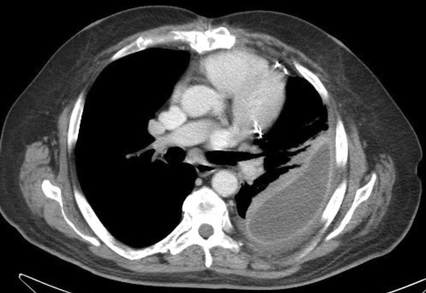
And metastases in the left midhemithorax.
Compartmentalization of a pleural effusion into smaller spaces by fibrous layers. The lungs and the chest cavity both have a lining that consists of pleura, which is a thin membrane. They may result from a variety of pathological processes which overwhelm the pleura's ability to reabsorb fluid. Differentiation of loculated effusions from solid. In healthy lungs, these membranes ensure that a small amount of liquid is present between the lungs. Diffuse nodules and opacification in right lung with compressive. Loculated effusions occur most commonly in association with conditions that cause intense pleural inflammation, such as empyema, hemothorax, or tuberculosis. The effusion may cause you to become breathless. Malignant pleural effusions (mpe) are the accumulation of pleural fluid and cancerous cells within the pleural space, occurring from neoplastic coronal cect of the same patient shows a large loculated left pleural effusion with circumferential pleural thickening. There is normally a tiny amount of fluid between the two layers of pleura. Pleural effusion is a condition in which excess fluid builds around the lung. A pleural effusion is a buildup of fluid between the layers of tissue that line the lungs and chest cavity. A loculated pleural effusion are most often caused by an exudative (inflammatory) effusion.
The pleura is a thin membrane that lines the inside of the chest wall and covers the lungs loculated pleural effusion. Exudate pleural effusion, hydrostatic pressure, inflammation, pleural effusion, transudate pleural effusion.